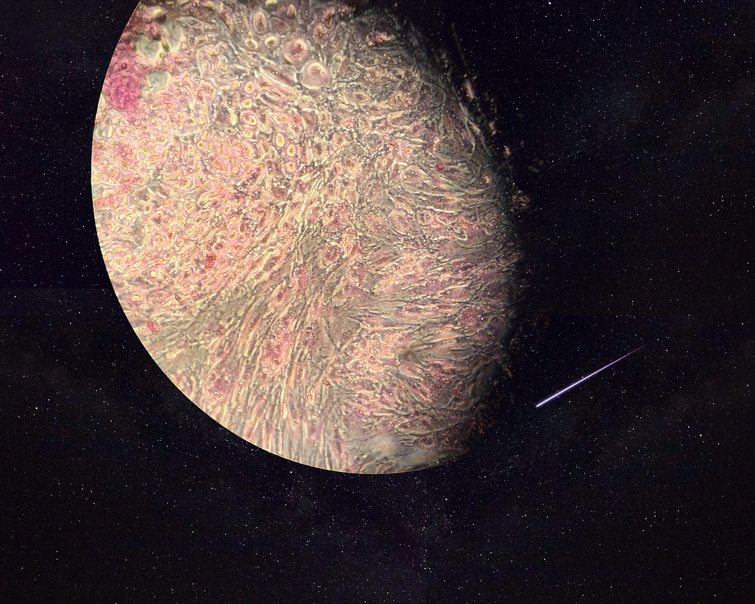
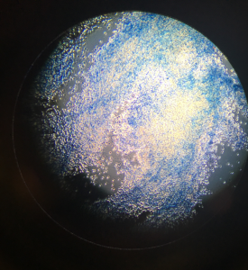
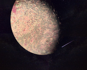
The image is shown through a microscopic view of endothelial cells that have been trypsinized. The image is portrayed as Earth, referring to it as a world for stem cells since endothelial cells can be used for the growth of stem cells.
Neurons, differentiated from human embryonic stem cells, stained with DAPI, beta-TubulinIII (white) and Ki67 (red), remind me of the classic movie The Matrix. “The Matrix is everywhere. It is all around us. Even now, in this very room. You can see it when you out your window or when you turn on yo...
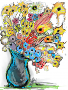
The sketch represents how I see a stems cells' potential--- a single cells can develop " bloom " and differentiate into a variety of types of cells. The base of the vase represents the pluripotent stem cells and the cells that it differentiates into such as other precursors, neuron, blood, intestina...
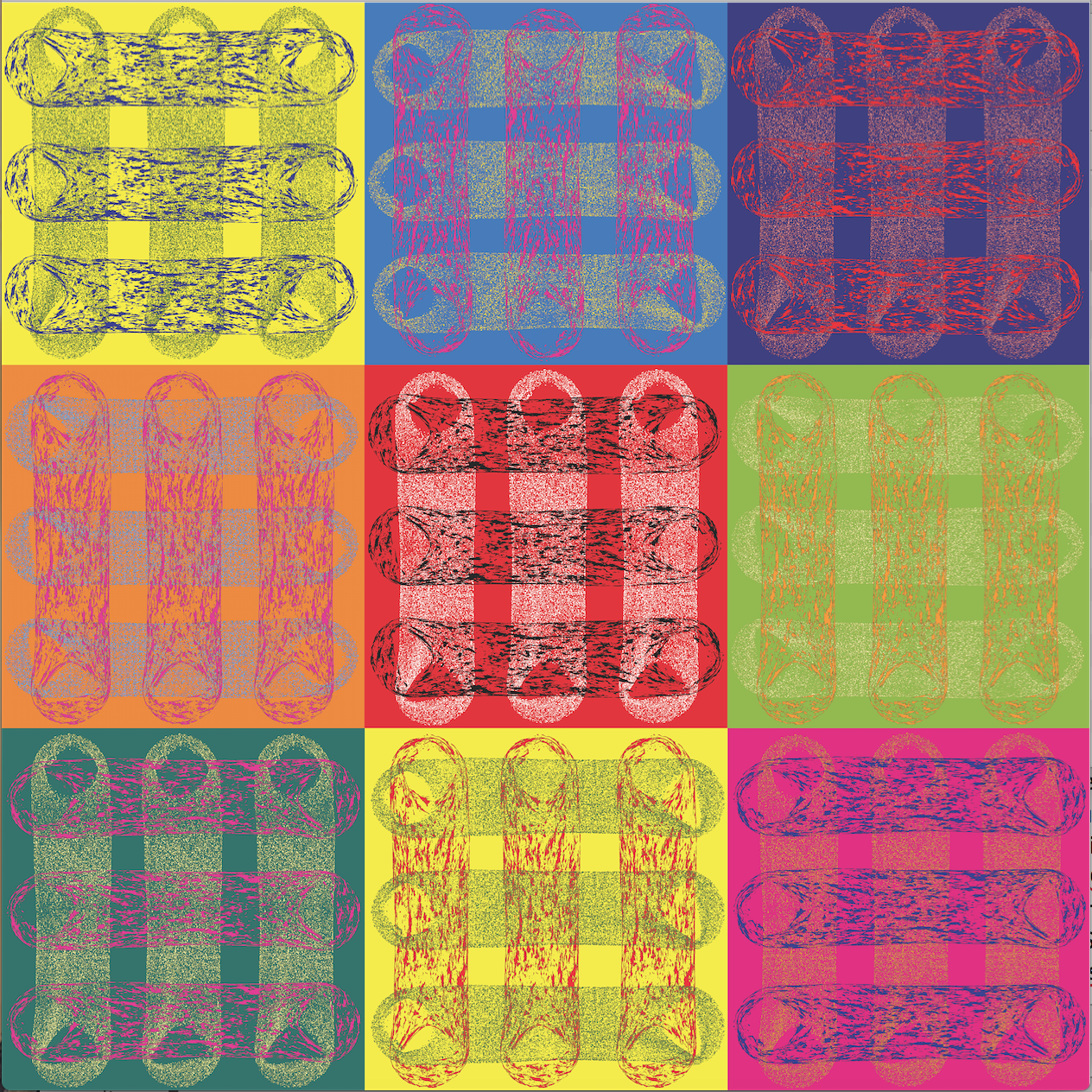
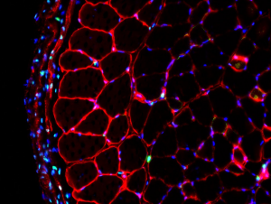
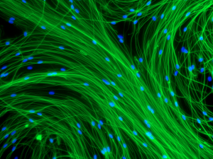
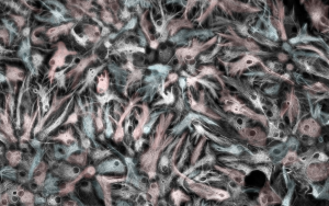
Inspired by Andy Warhol’s artwork depicted in this photo is an array of skeletal muscle tissue fibroblasts and nuclei arranged in diverse colours.
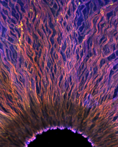
A transverse section of a skeletal muscle imaged with a confocal microscope.
Here we show a GFP-positive newly formed ensheathing/myelinating oligodendrocyte observed after a contusion spinal cord injury in a PDGFRaCreER: Rosa26-mGFP(mT/mG) transgenic mouse spinal cord. Our lab recently showed that PDGFRa+ oligodendrocyte precursor cells (OPCs) can form multiple different gl...
There is always a light at the end of the tunnel… keep dreaming!
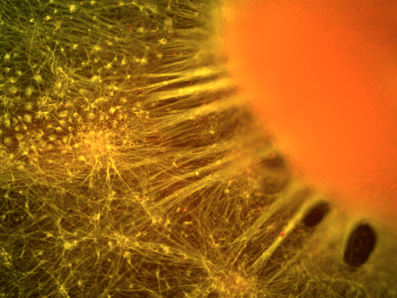
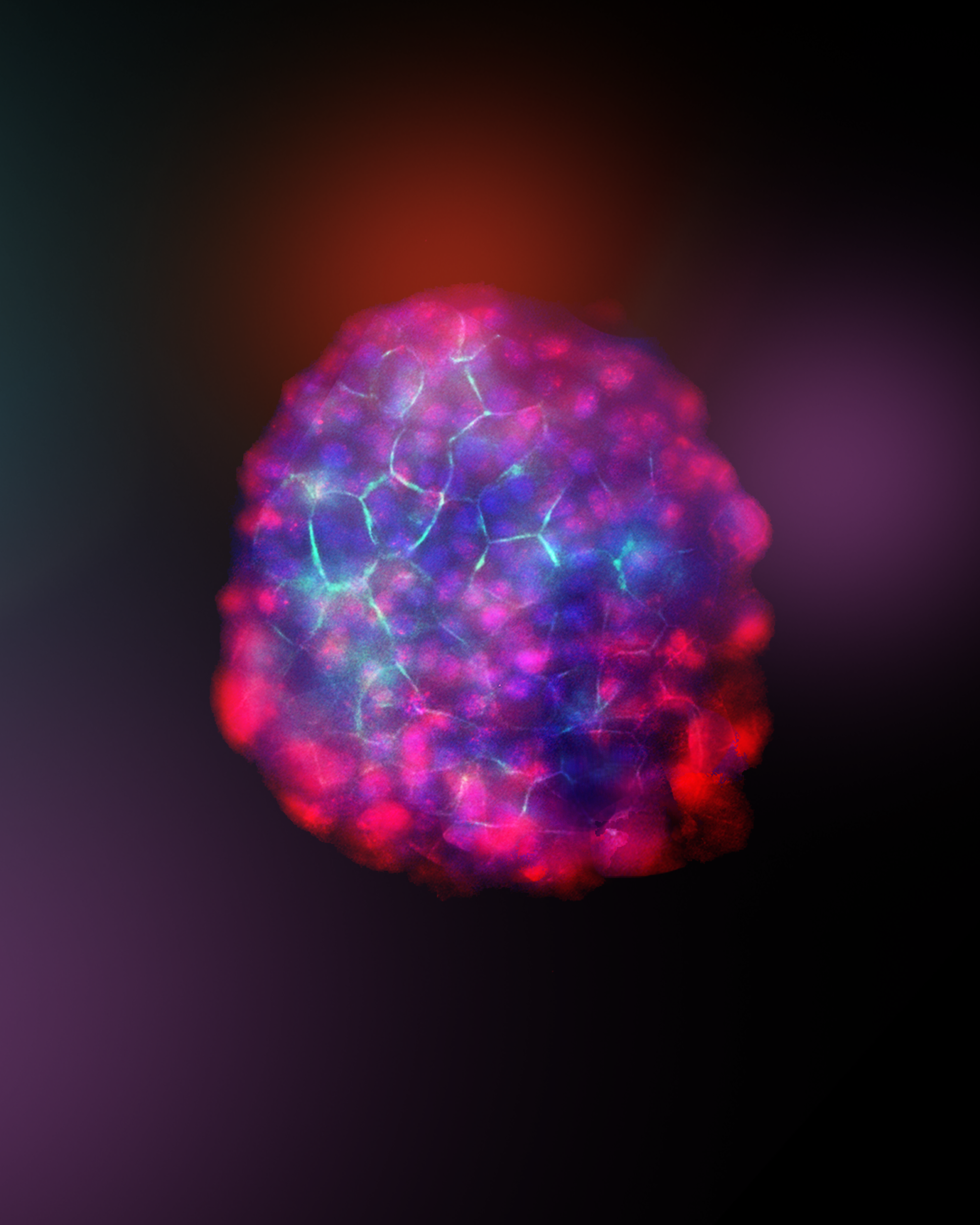
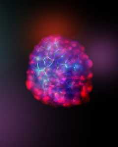
This jellyfish-like structure is composed of a neural aggregate differentiated from human induced pluripotent stem cells with drug releasing microparticles. The tentacle-like projections of the neurites growing and reaching out to form connections resemble a jellyfish swimming in the ocean.
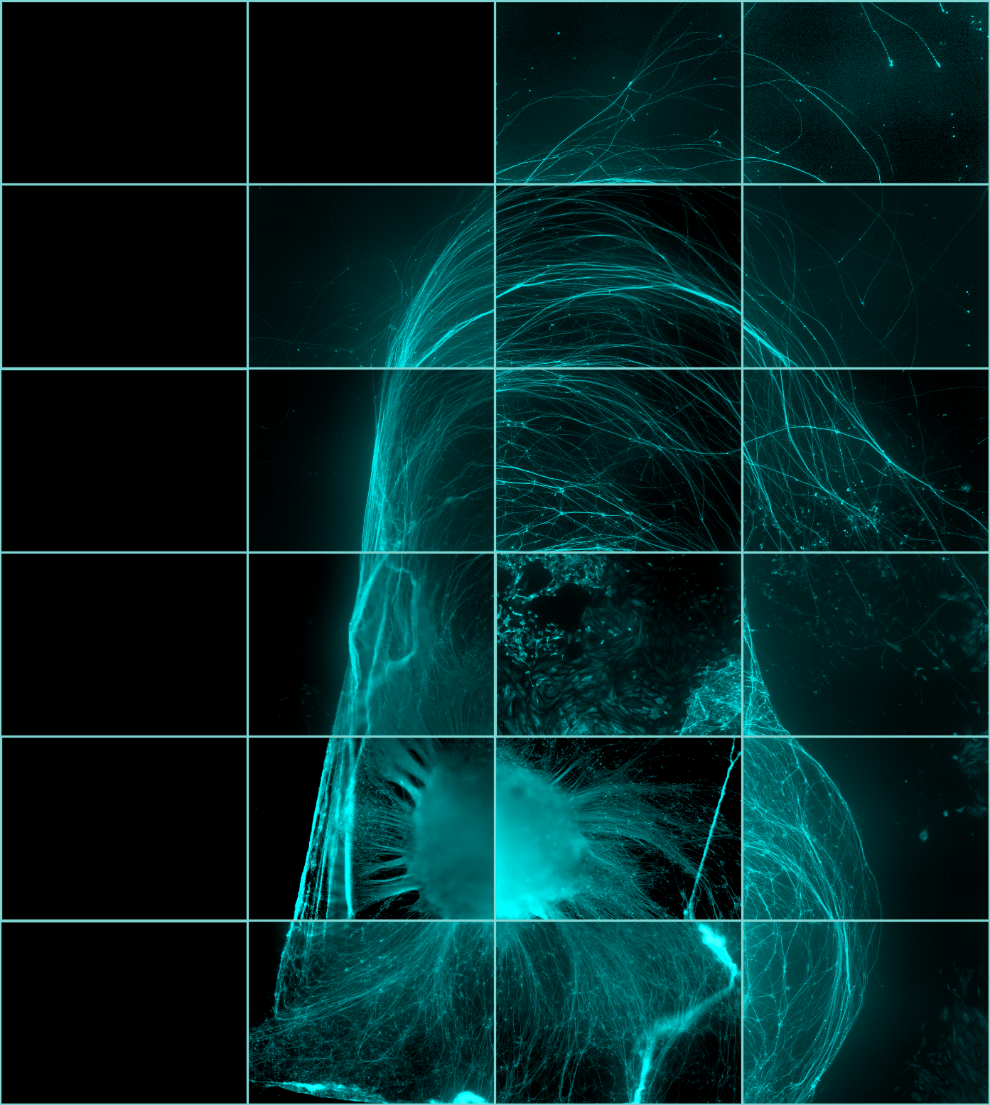
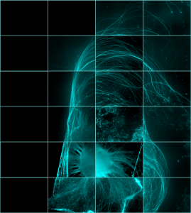
Axonal threads fling themselves from the neural aggregate like flares from the sun. Human induced pluripotent stem cells were differentiated into neural tissue.
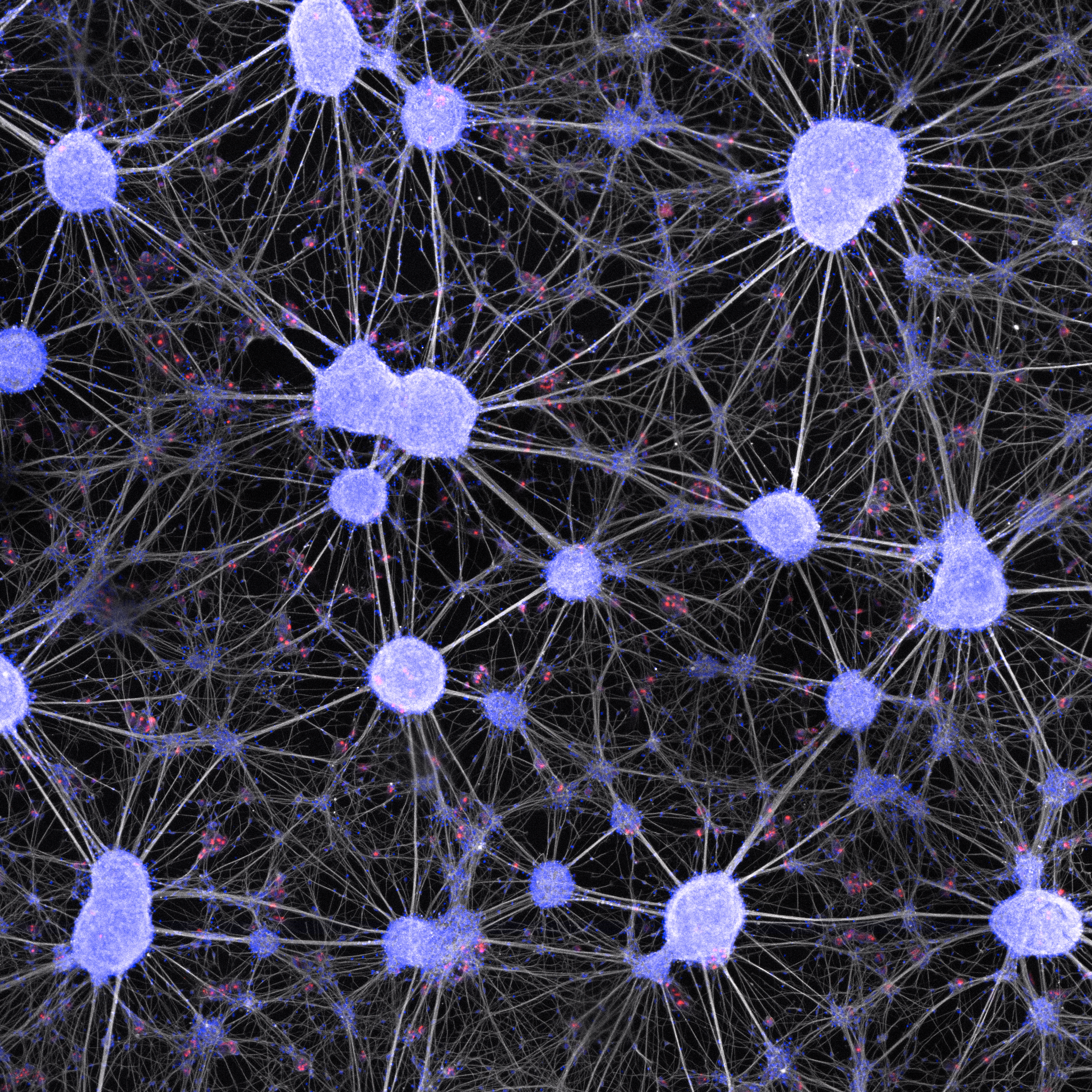
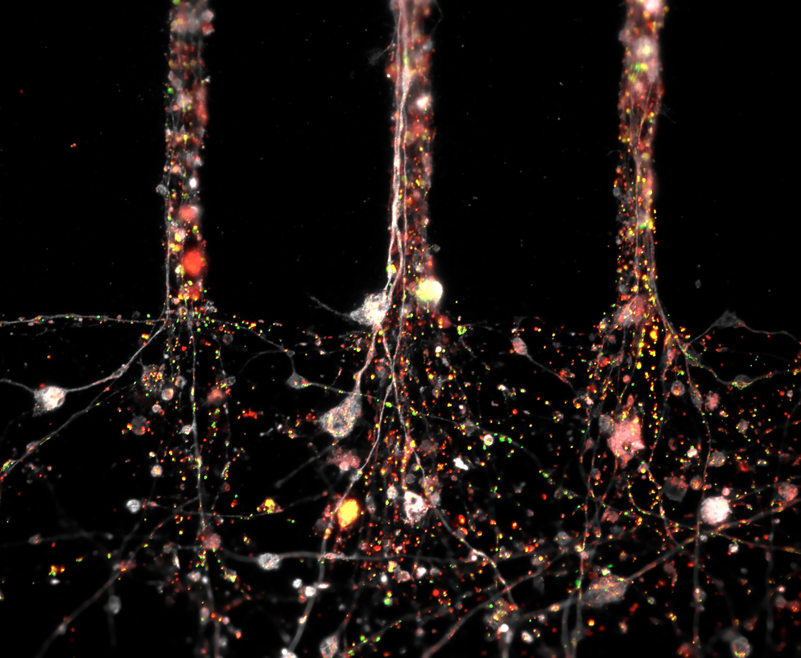
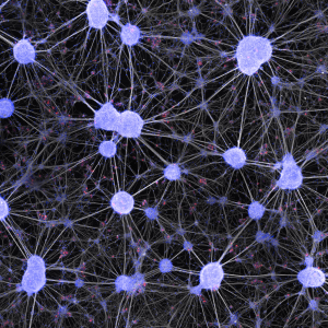
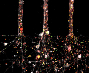
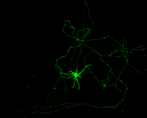
A Neuronal Network in a Dish: These neurons have been formed by the differentiation of adult human spinal cord neural cells, which represent a promising endogenous target for treating spinal cord disorders.
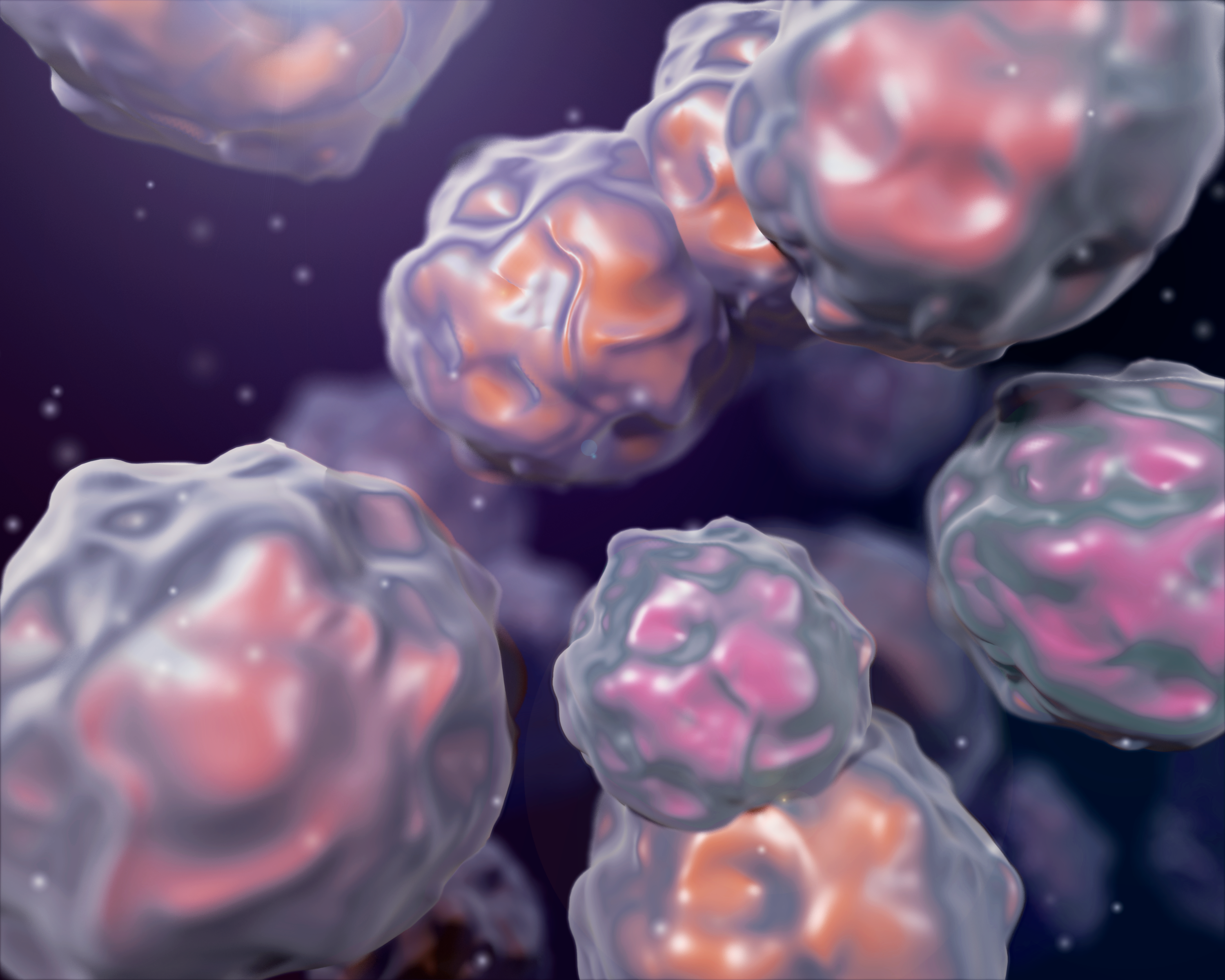
3D illustration inspired by SEM images of stem cells with a cinematic flare.
Human dopaminergic neurons differentiated from induced pluripotent stem cells in the microfluidics devices created by the company ANANDA Devices.
Image of neural progenitor cells that spontaneously differentiated into neurons (Tuj1+) in yellow and oligodendrocytes (O4+) in red. Cells appear as supernovas, dancing and dazzling in the night sky.
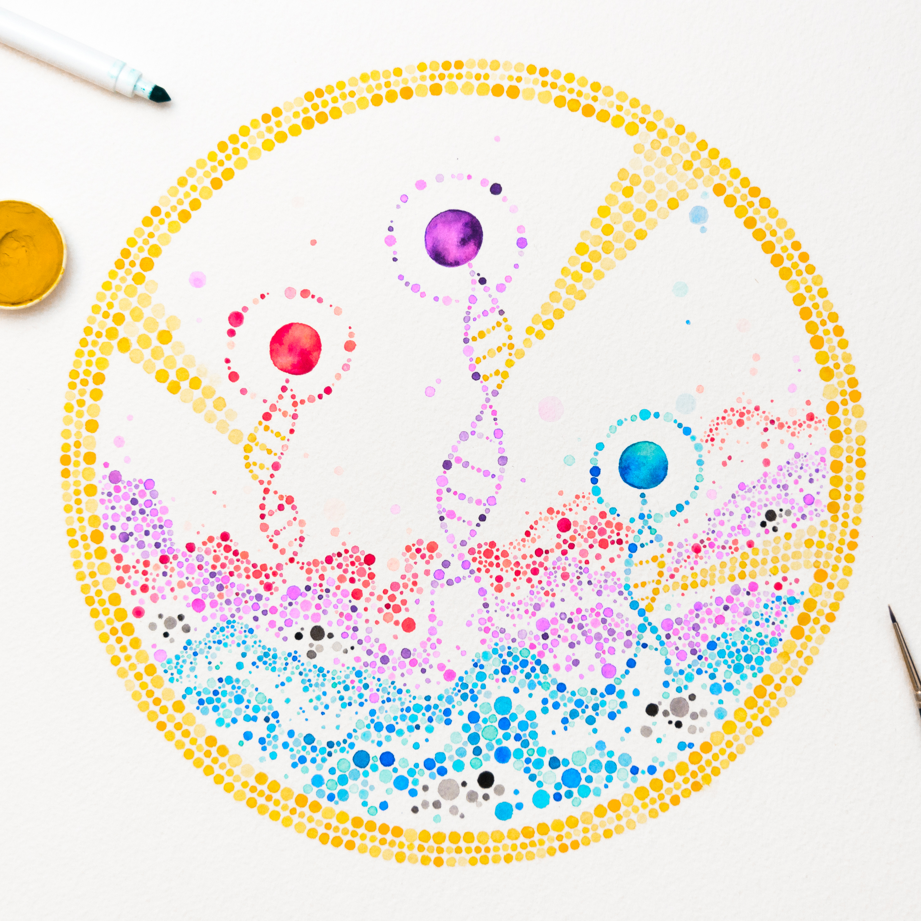
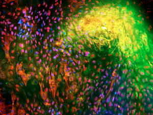
Cells are constantly competing with one another for nutrients and space. Sometimes, "winner" cells will directly eliminate their less-fit ("loser") neighbours. As a result of competition, elite cells may take over the population. In this watercolour piece, pointillism is used to capture competition ...
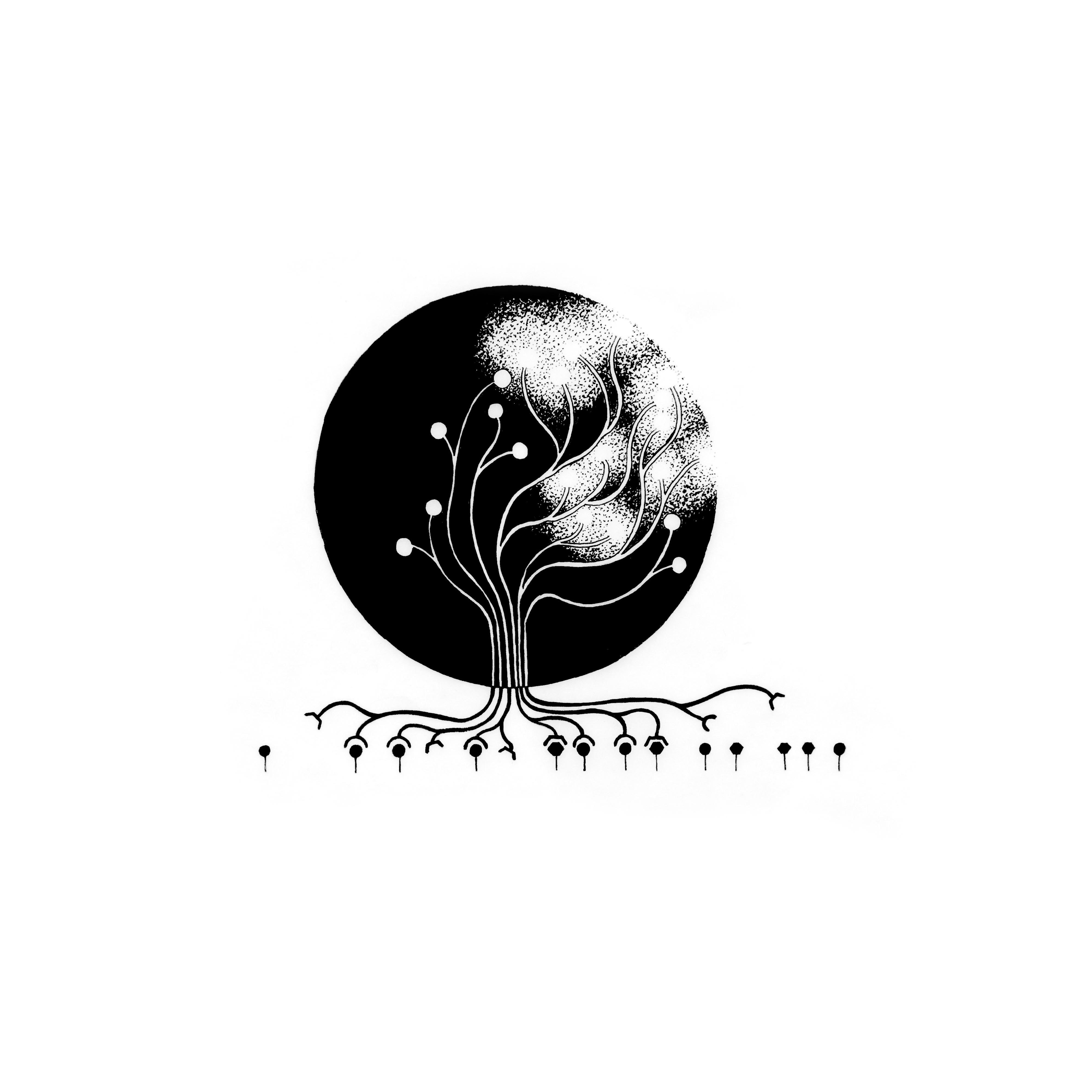
Much like a growing tree, cells can sense their environment & let it guide their growth & fate. This hand-drawn illustration represents how synergistic signaling can trigger cascades of regulatory events and impact T cell lineage commitment. The Tree of Fate connects the endogenous genetic p...
Glycogen storage revealed by Period acid-Schiff staining in IPSCs-derived hepatocytes.
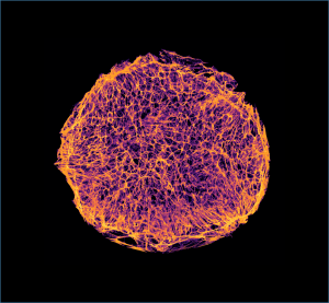
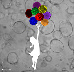
“One image is worth a thousand words.” This image represents the beauty of the 3D cell culture.
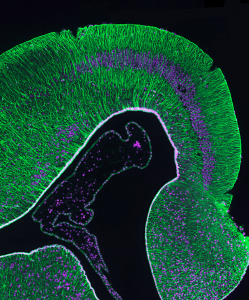
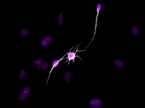
Radial glial are a type of stem cell which have long processes spanning the brain. Radial glia rise to new brain cells, which use the processes as a path to travel to their final destination. Though they are not present in all adult animals, reptiles such as the gecko (studied in our lab), keep them...
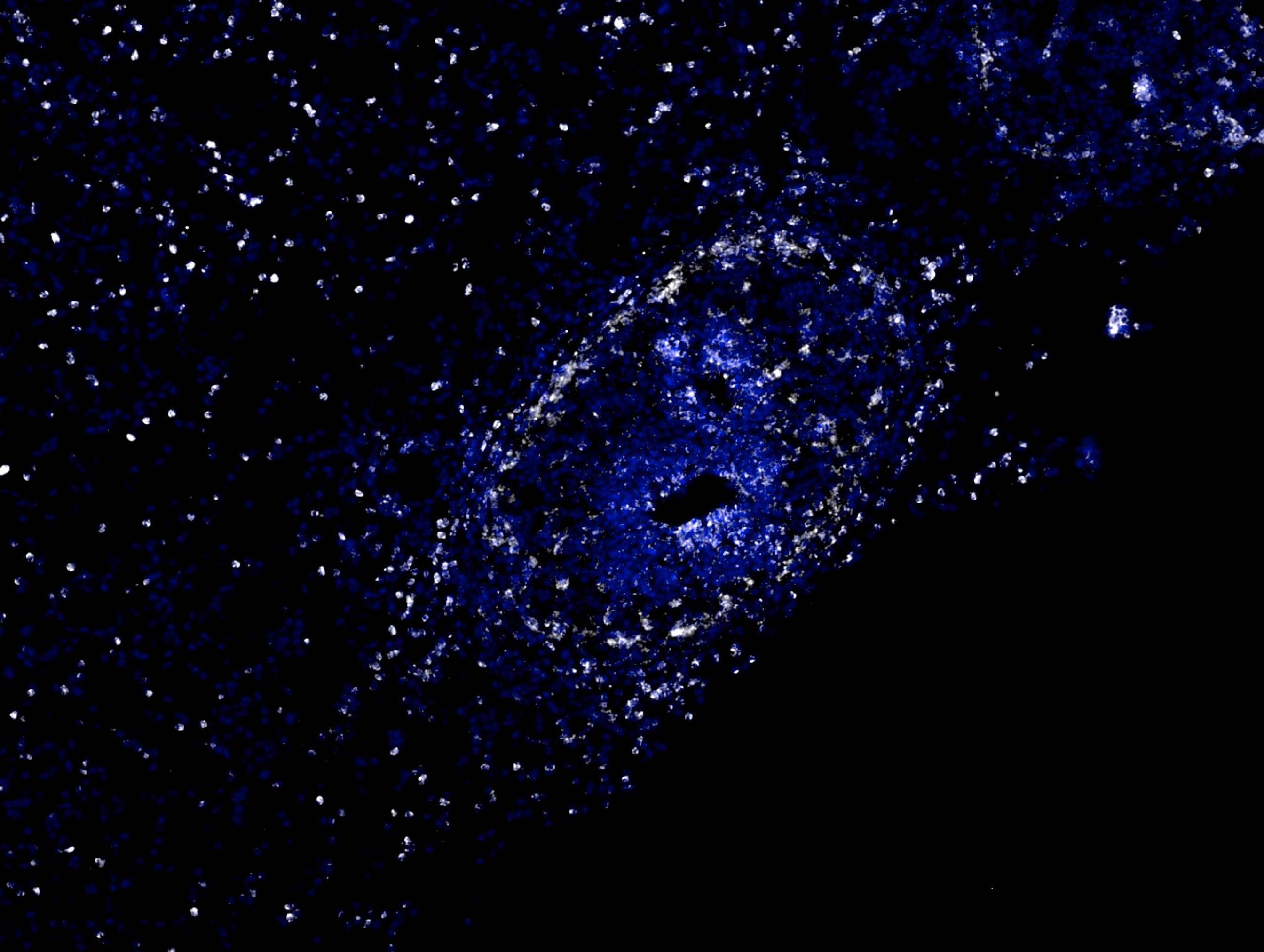
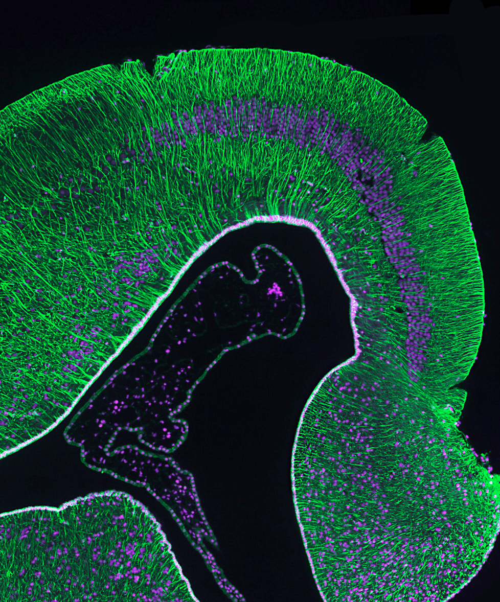
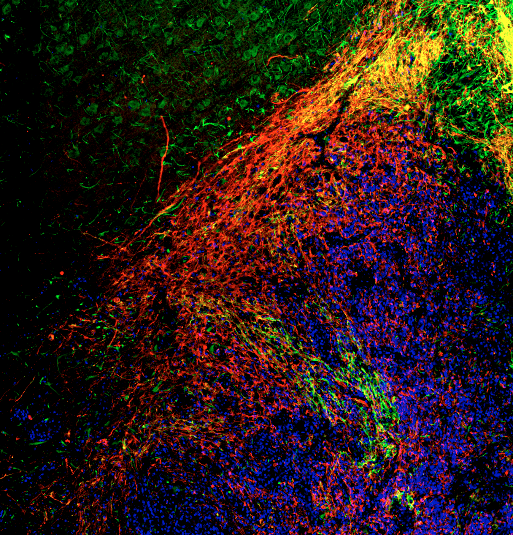
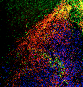
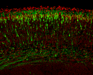
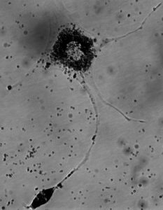
This image depicts a rat brain that has been transplanted with human neural stem cells. Human cells are coloured red, and migrating cells (both rat and human) are coloured green. Overlap between red and green (yellow) represents migrating human cells. In this image I see migrating human cells sweepi...
Human umbilical cord derived stem cells differentiating into cardiomyocytes. Stainings: human nuclear antigene, sarcomeric actinin, SERCA, DAPI.
Human umbilical cord derived stem cells differentiation into cardiomycytes. Stainings: human nuclear antigene, sarcomeric actinin, SERCA, DAPI.
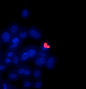
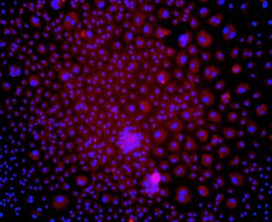
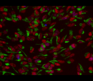
The differentiation of induced pluripotent stem cells into a more mature phenotype is not 100% efficient. Here were see the evolution of two distinct cell types form the same culture.
Some people perform better when under pressure. Likewise, some cells thrive when subjected to microscale forces. Here, we are studying the effect of tensional forces on induced pluripotent stem cell differentiation by micropatterning culture surfaces.
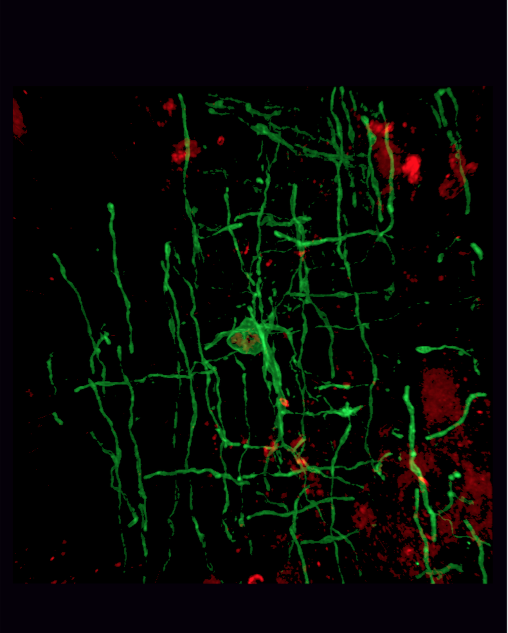
Like rubber around an electric wire, the cells in pink here insulate the electric message-conducting neurons in your brain, letting them send messages faster and more efficiency. They are called “oligodendrocytes,” which translates to “cells with many branches,” and they’re some of the las...
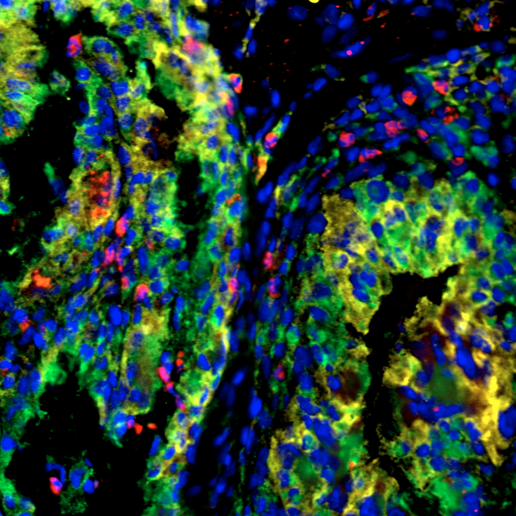
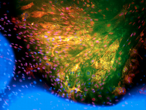
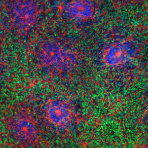
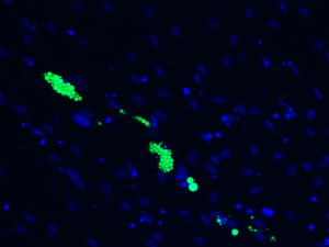
Macrophages (CD68+, green) express arginase-1 (Arg-1, yellow) when infiltrating an intramuscularly injected hydrogel containing rat adipose-derived stem/stromal cells (red). DAPI counterstain (blue).