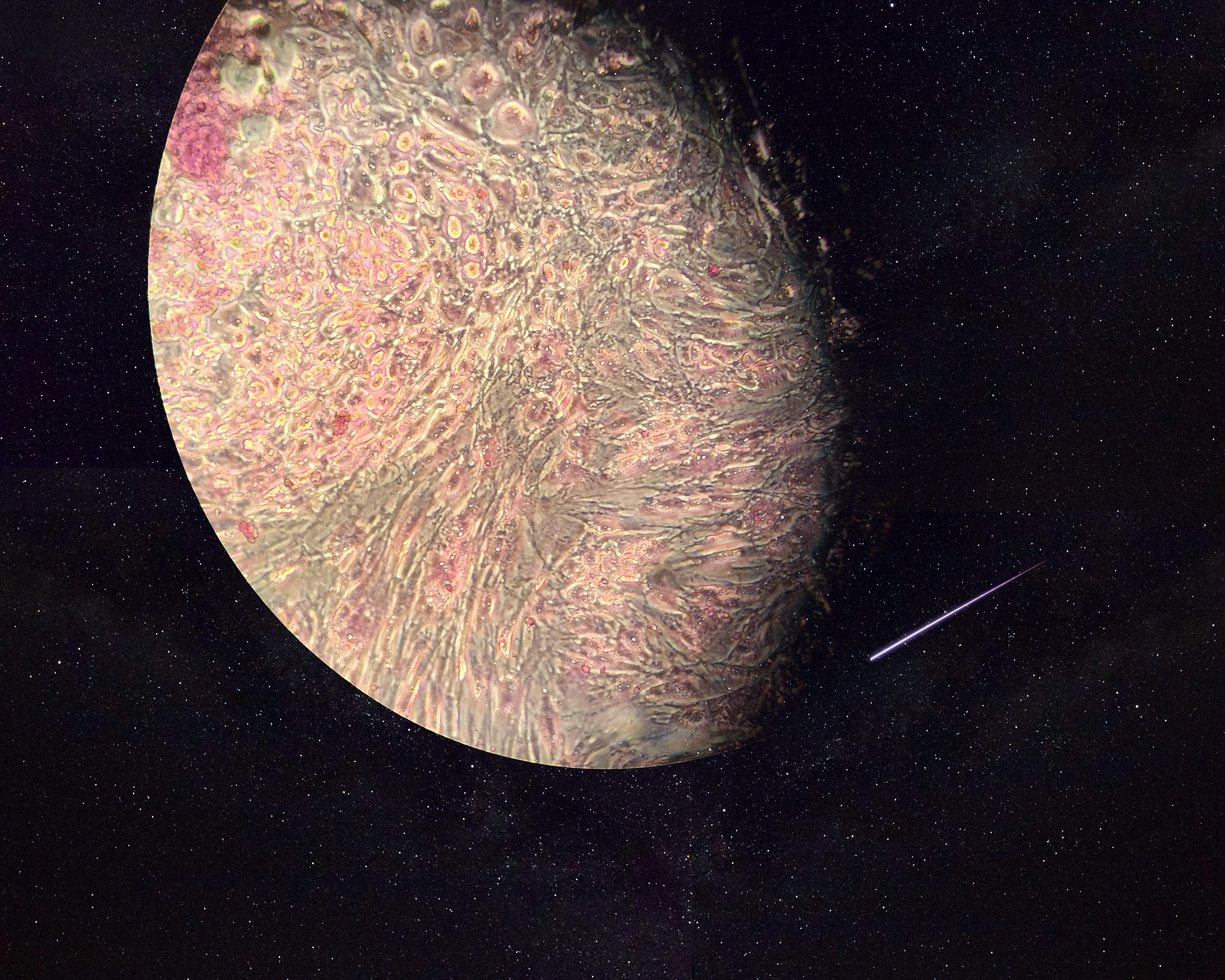
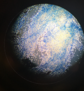
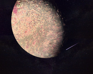
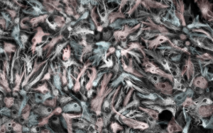
The image is shown through a microscopic view of endothelial cells that have been trypsinized. The image is portrayed as Earth, referring to it as a world for stem cells since endothelial cells can be used for the growth of stem cells.
Neurons, differentiated from human embryonic stem cells, stained with DAPI, beta-TubulinIII (white) and Ki67 (red), remind me of the classic movie The Matrix. “The Matrix is everywhere. It is all around us. Even now, in this very room. You can see it when you out your window or when you turn on yo...
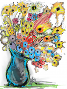
The sketch represents how I see a stems cells' potential--- a single cells can develop " bloom " and differentiate into a variety of types of cells. The base of the vase represents the pluripotent stem cells and the cells that it differentiates into such as other precursors, neuron, blood, intestina...
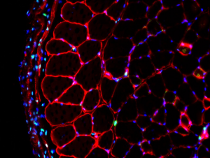
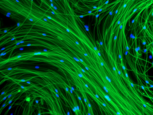
Inspired by Andy Warhol’s artwork depicted in this photo is an array of skeletal muscle tissue fibroblasts and nuclei arranged in diverse colours.
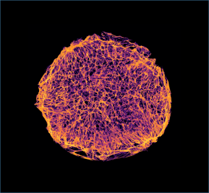
A transverse section of a skeletal muscle imaged with a confocal microscope.
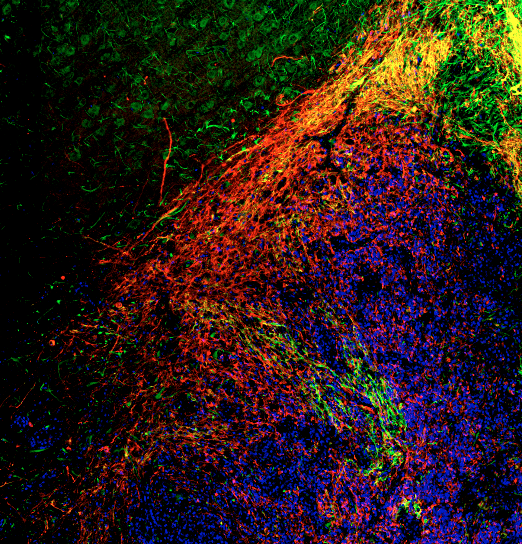
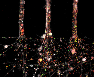
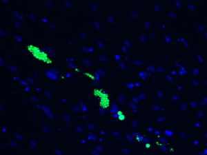
Here we show a GFP-positive newly formed ensheathing/myelinating oligodendrocyte observed after a contusion spinal cord injury in a PDGFRaCreER: Rosa26-mGFP(mT/mG) transgenic mouse spinal cord. Our lab recently showed that PDGFRa+ oligodendrocyte precursor cells (OPCs) can form multiple different gl...
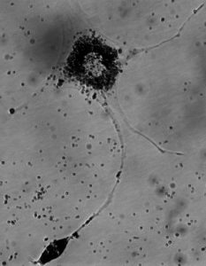
There is always a light at the end of the tunnel… keep dreaming!
This jellyfish-like structure is composed of a neural aggregate differentiated from human induced pluripotent stem cells with drug releasing microparticles. The tentacle-like projections of the neurites growing and reaching out to form connections resemble a jellyfish swimming in the ocean.
Axonal threads fling themselves from the neural aggregate like flares from the sun. Human induced pluripotent stem cells were differentiated into neural tissue.
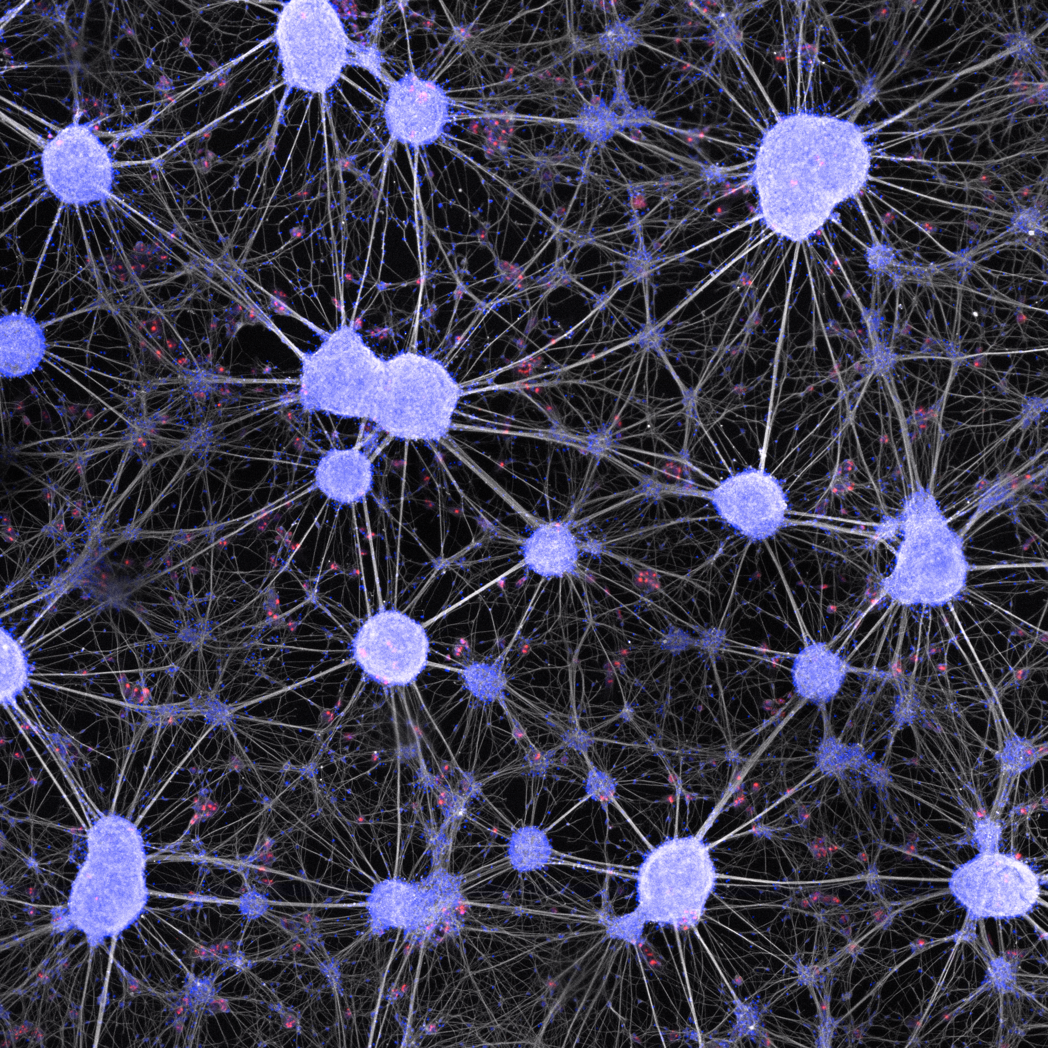
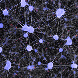
A Neuronal Network in a Dish: These neurons have been formed by the differentiation of adult human spinal cord neural cells, which represent a promising endogenous target for treating spinal cord disorders.
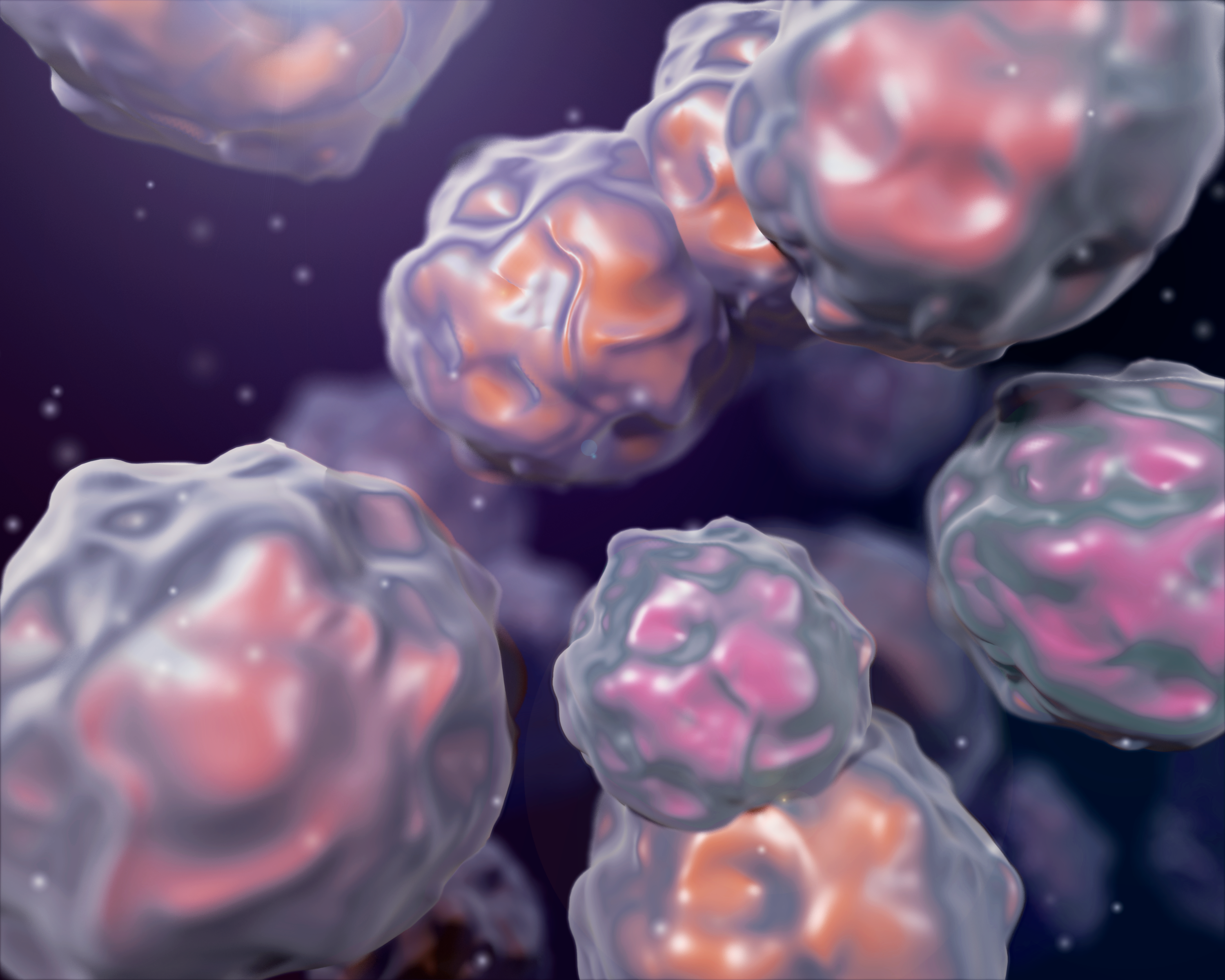
3D illustration inspired by SEM images of stem cells with a cinematic flare.
Human dopaminergic neurons differentiated from induced pluripotent stem cells in the microfluidics devices created by the company ANANDA Devices.
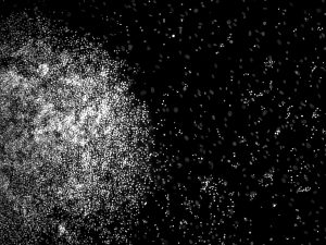
Image of neural progenitor cells that spontaneously differentiated into neurons (Tuj1+) in yellow and oligodendrocytes (O4+) in red. Cells appear as supernovas, dancing and dazzling in the night sky.
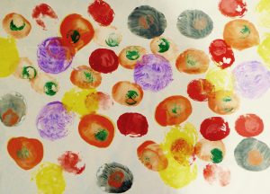
Cells are constantly competing with one another for nutrients and space. Sometimes, "winner" cells will directly eliminate their less-fit ("loser") neighbours. As a result of competition, elite cells may take over the population. In this watercolour piece, pointillism is used to capture competition ...
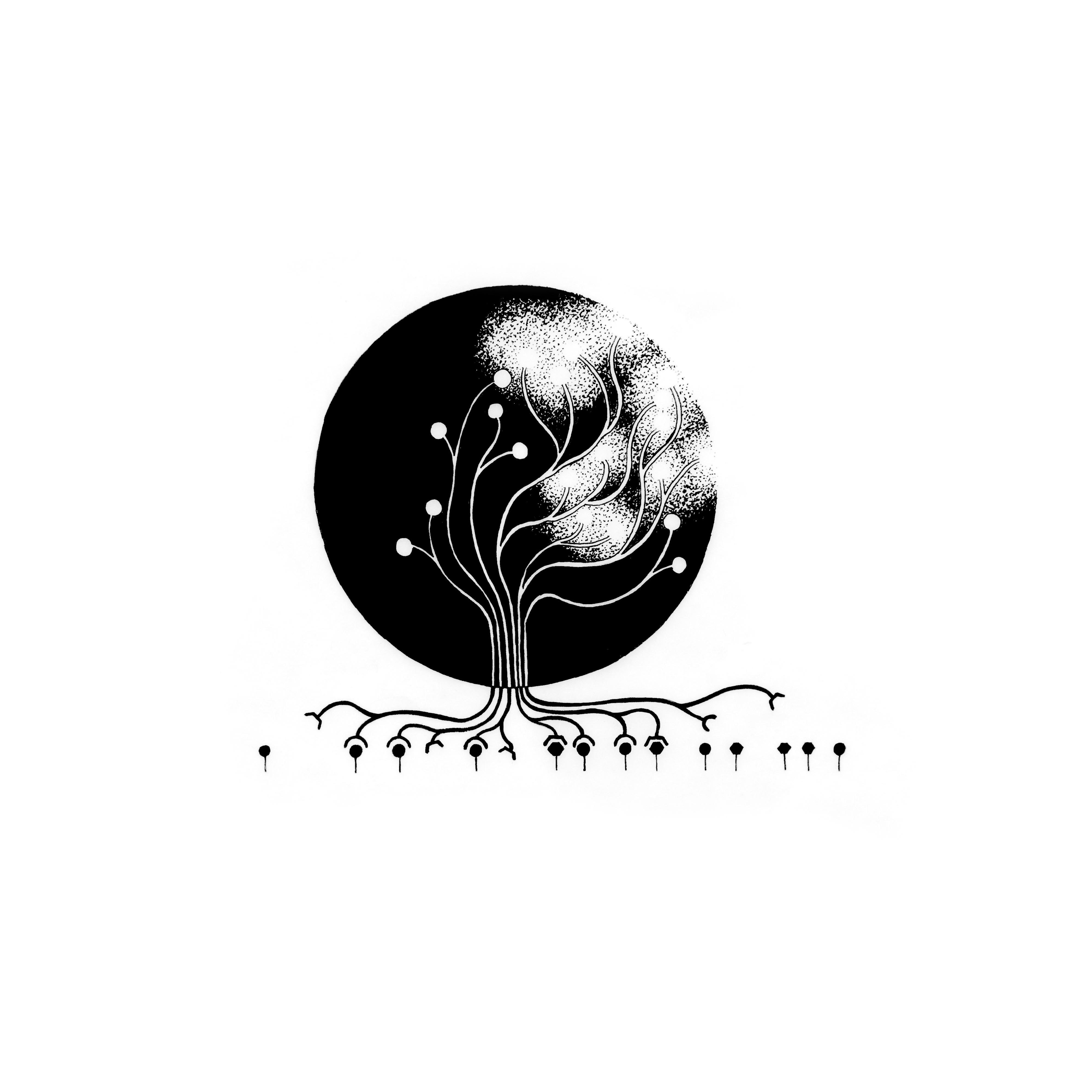
Much like a growing tree, cells can sense their environment & let it guide their growth & fate. This hand-drawn illustration represents how synergistic signaling can trigger cascades of regulatory events and impact T cell lineage commitment. The Tree of Fate connects the endogenous genetic p...
Glycogen storage revealed by Period acid-Schiff staining in IPSCs-derived hepatocytes.
“One image is worth a thousand words.” This image represents the beauty of the 3D cell culture.
Radial glial are a type of stem cell which have long processes spanning the brain. Radial glia rise to new brain cells, which use the processes as a path to travel to their final destination. Though they are not present in all adult animals, reptiles such as the gecko (studied in our lab), keep them...
This image depicts a rat brain that has been transplanted with human neural stem cells. Human cells are coloured red, and migrating cells (both rat and human) are coloured green. Overlap between red and green (yellow) represents migrating human cells. In this image I see migrating human cells sweepi...
Human umbilical cord derived stem cells differentiating into cardiomyocytes. Stainings: human nuclear antigene, sarcomeric actinin, SERCA, DAPI.
Human umbilical cord derived stem cells differentiation into cardiomycytes. Stainings: human nuclear antigene, sarcomeric actinin, SERCA, DAPI.
The differentiation of induced pluripotent stem cells into a more mature phenotype is not 100% efficient. Here were see the evolution of two distinct cell types form the same culture.
Some people perform better when under pressure. Likewise, some cells thrive when subjected to microscale forces. Here, we are studying the effect of tensional forces on induced pluripotent stem cell differentiation by micropatterning culture surfaces.
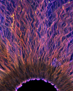
Like rubber around an electric wire, the cells in pink here insulate the electric message-conducting neurons in your brain, letting them send messages faster and more efficiency. They are called “oligodendrocytes,” which translates to “cells with many branches,” and they’re some of the las...
Macrophages (CD68+, green) express arginase-1 (Arg-1, yellow) when infiltrating an intramuscularly injected hydrogel containing rat adipose-derived stem/stromal cells (red). DAPI counterstain (blue).

 and SOX2 (a nuclear marker shown in yellow). All nuclei are stained with DAPI (represented in purple). Photo: E600 microscope.")
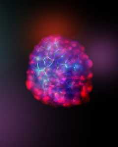
 is a hematological malignancy. AML has a poor prognosis rate, driven and perpetuated by leukemia stem cells (LSCs). Mitochondrial homeostasis has been demonstrated as an effective target to eradicate LSCs. However, the mood of mitochondria to LSC function requires further investigation. We hypothesize that mitochondrial mood is essential for LSC survival. We performed transmission electron microscopy (TEM) imaging on AML cells to interrogate this question and observed just one angry mitochondrion (Figure 1). The data suggests that the AML cells can keep mitochondria happy to increase oxidative phosphorylation (OXPHOS). OXPHOS is a metabolic process in the mitochondria and has been reported to be crucial for LSC survival. Altogether, our preliminary data infers the importance of mitochondrial mood to AML cells. Although this hypothesis requires further evidence, we hope this image makes your day.")

 and the journey from one to the many (differentiation).")

, and the nuclei are stained by DAPI (in blue).")

, LDHA (red), and SOX2 (green). Image taken on a Nikon Ti2e microscope. Z-stacks were processed in NIS-Elements AR using Clarify.ai and Extended Depth of Focus.")
, while the core may retain some pluripotent cells (stained in pink). These residual pluripotent cells can pose risks in cell therapies, as they are not fully differentiated. However, in the regular culture of iPSCs, this mix of cell types can be seen as a natural balance, much like the concept of yin and yang, where different elements coexist and influence each other.")

 and A549 cancer cells (red mPLUM) were seeded to form a 3D spheroid. Unexpectedly, they shaped a heart, demonstrating an unplanned interaction between the cell types in a unique, bioluminescent structure. The image was captured and analyzed using the Incucyte S3 Live-Cell Analysis System.")
. The dendrites are depicted in red, marked by the MAP2 antibody, while the nuclei of cells are shown in blue (DAPI).")
 captures a vivid snapshot of the battlefield. The vibrant pancreatic islets glow in radiant hues of yellow and red, with the golden clusters representing the precious beta cells. But the real drama unfolds as immune cells—marked in striking blue and green—mount an aggressive assault, infiltrating the islets, and waging an attack on the beta cells, captured in the pancreas of a mouse as it develops type 1 diabetes.")
.")
 on a decellularized adipose tissue biomaterial (blue). Where the biomaterials came in contact created areas void of cells producing this spooky fellow.")
 stained with specific maturity markers.")
 were stained with multiple specific markers.")
, Perilipin-1 (red), and DAPI (blue). The image was acquired using an LSM 900 Zeiss microscope.")


 activate and become proliferating myoblasts. These myogenic progenitors eventually exit the cell cycle to differentiate into myocytes. Their fusion leads to the generation of multinucleated myotubes. This is followed by the maturation of the myotubes which form myofibers: the main component of the skeletal muscles. The phenotypic fate (slow or fast) of newly generated muscle fibers is influenced by the intrinsic programming of the satellite cells and complex extrinsic signals (e.g., innervation).")
 derived from something as simple and as trivial as urine to create beautiful bouquets of iPSCs to study severe neurodevelopmental diseases. In this image, iPSC markers SSEA4 (green), OCT4 (red), and DAPI (blue) are fluorescently labeled to verify the presence of stem cell markers with the objective of further differentiating these cells into a variety of neural cell types such as neural progenitor cells, astrocytes, oligodendrocytes, and cortical neurons.")
 labeled with marker O4 (cyan, left), and microglia in a colony, labeled with Iba-1 (purple, right) that are co-stained with the nuclear marker DAPI (yellow). These two distinct cell types were derived from the same in vitro culture, and the scene is reminiscent of another fascinating interaction that occurs in the depths of the ocean. The OPC on the left is strikingly like the luminescent fin of the angler fish, which acts as a lure to draw in other fish; whereas the morphology of microglia mimics those of small fish, traveling as a school or colony. However, in the brain, the roles are reversed, with microglia secreting factors that promote OPC migration. This piece is inspired by the notion that the development of two completely different cells that have entirely unique morphologies and functions and are derived from distinct lineages can be supported by the same environments. A parallel is drawn between these cells and two unique species like anglerfish and their prey, which also serve different functions and have distinct morphologies, but are supported by the deep sea. Together these different living creatures function as part of an ecosystem, just as OPCs and microglia function together to serve an indispensable role in the central nervous system.")
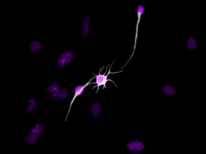
 project their axons from small E14.5 mouse retinal explants. This 24-hour culture is called RGC outgrowth assay. The RGC’s neurite is labeled with neurofilament (2H3) antibody in red and the nuclei with Hoechst in blue.")
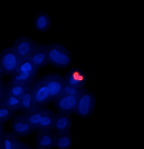
 shows cells are in the proliferation stage.
Red is rhodamine-phalloidin binds to actin and blue is DNA.")
. The neural stem cells pictured here have a severe mutation that causes a rare and fatal neurodevelopmental disorder. Using organelle-specific fluorescent dyes, we can observe extremely aberrant structures within the cells. The image shows nuclei (blue), lysosomes (magenta), and mitochondria (yellow). Culturing and differentiating these mutant neural stem cells into neurons will help us understand the development of the disease and ways to treat it.")
 clinging on to the adjacent blood vessel (magenta) in an ‘awkward hug’ in the zebrafish trunk. These cells provide critical support for the developing vasculature and function as progenitors for pericytes and tendon fibroblasts post-injury.")
 depositing collagen protein fibrils (green) around a single blood vessel in the zebrafish trunk.")

 is a useful tool to study brain development. This picture shows an organoid that was cut after 47 days of differentiation. Growing from the inside and branching out towards the outer regions, the cells within this “mini-brain” represent different cell populations, where the less differentiated cells can be found in the inner part of the structure (stained in turquoise with neural stem cell marker SOX2). The differentiated cells are the outer ones (stained in magenta with mature neuronal marker MAP2), and the nucleus was stained with DAPI.")
 is a useful tool to study brain development. This picture shows an organoid that was cut after 25 days of differentiation: growing from the inside and branching out towards the outer regions, the cells within this “mini-brain” represent different cell populations, where the less differentiated cells can be found in the most inner part of each circle (stained in yellow with neural stem cell marker NESTIN) whereas the differentiated cells are the most outer ones (stained in red with mature neuronal marker MAP2). Since this is an early time point, there are not a lot of differentiated neurons. The nucleus was stained with DAPI.")
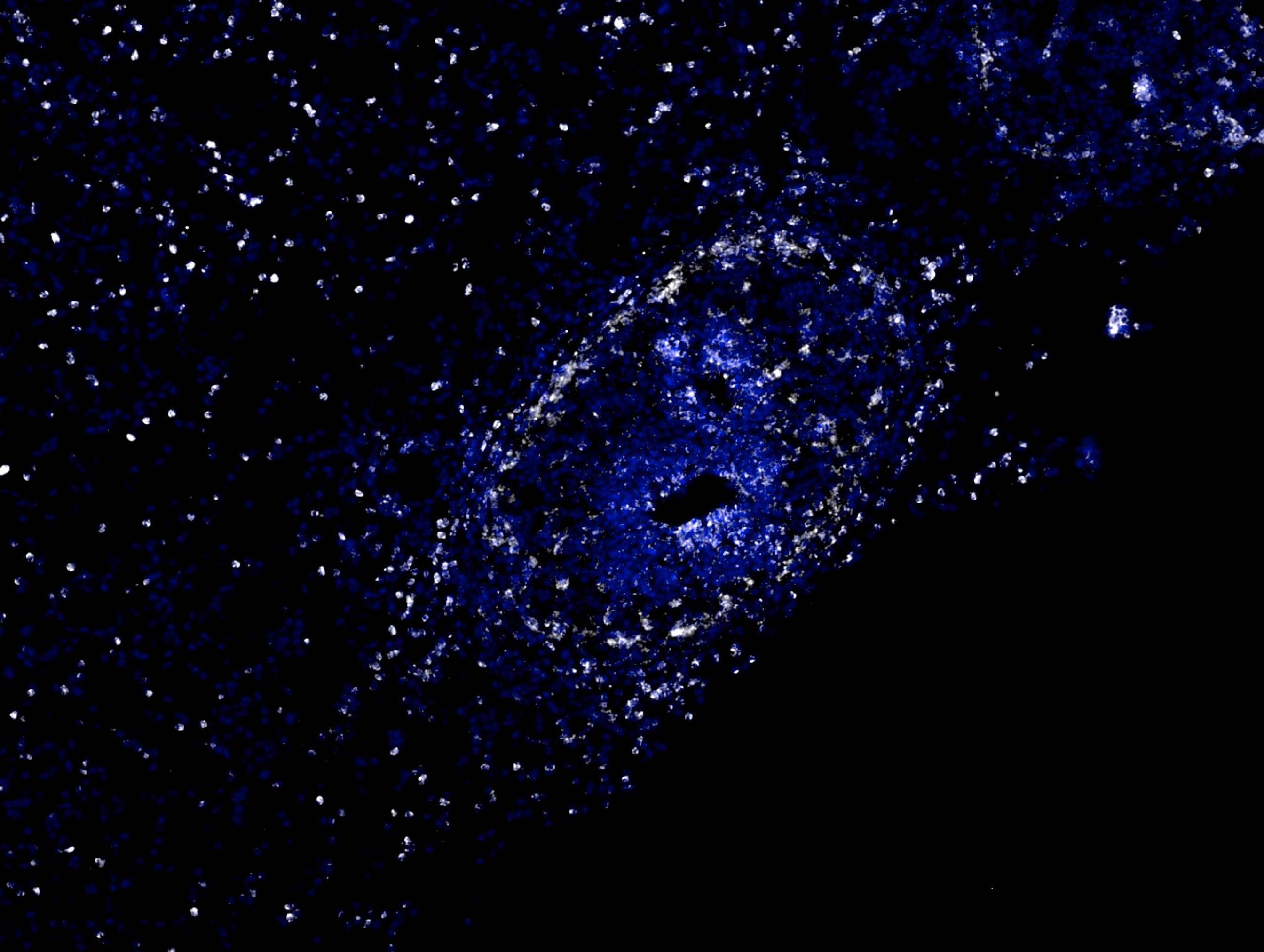
 mouse brain cut at a thickness of 8 uM. Staining for the stem cell marker, SOX2 (red), progenitor cell marker, TBR2 (green), and nuclei marker, DAPI (blue) was done by Dilan Rasool. Sectioning and scanning services were performed by the Cell Biology and Image Acquisition Core (RRID: SCR_021845) funded by the University of Ottawa, the Natural Sciences and Engineering Research Council of Canada, and the Canada Foundation for Innovation. The image was taken with a Zeiss Axioscan Fluorescence at 40X.")

, the white structures are vessels stained with lectin and the nuclei are blue. The red cell is coming from the Hic1-cre-tdTomato grafted piece of adipose tissue.")
, immune cells are in green (Cdk5) and nuclei are in blue (Hoechst). Aureus in the centre seems to be encapsulated by a fibroblast immune cell shell.")
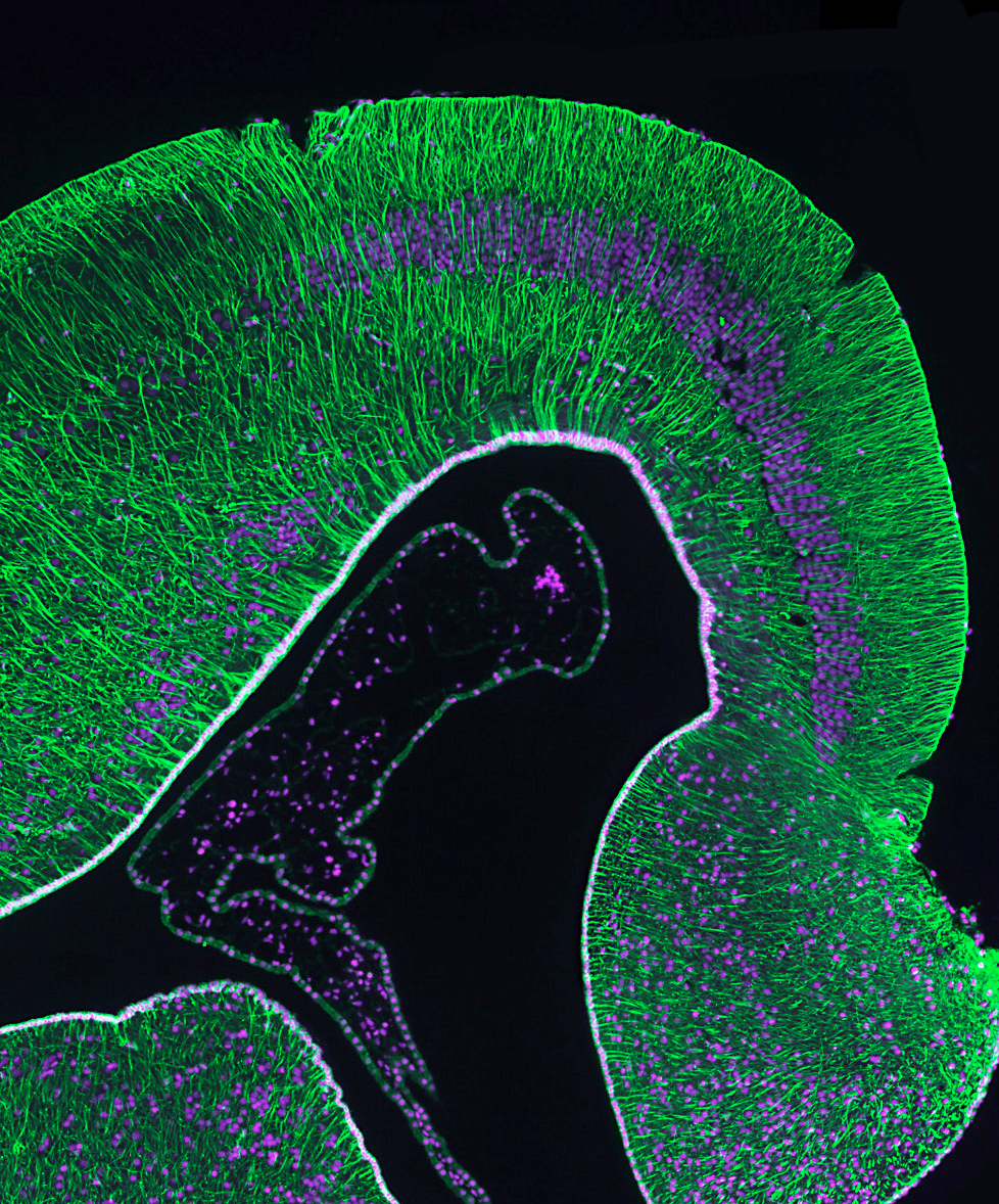
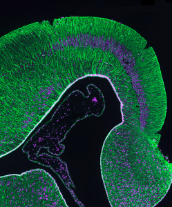
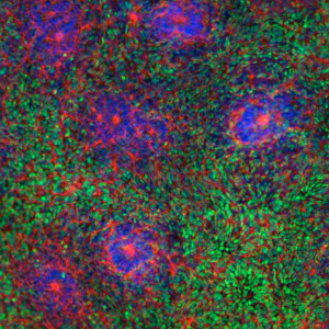
 which generate most of the neurons in the brain. At the periphery of the ventricles, intermediate progenitor cells (in red) actively proliferate and contribute to the greater size of the human brain. Layers of newly born neurons (in magenta) will form the cerebral cortex, which is the outermost part of the brain.")


, TUJI (red), and DAPI (blue), this cluster of neurons may resemble an organism trapped in a spider's web, but it has allowed us the freedom to explore human neurodevelopment in a dish.")
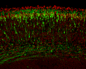
 have the surprising ability to self-organize into tissue structures by recapitulating certain aspects of embryonic development. We cultured hPSCs as aggregates in a neural differentiation media for 50 days to form brain organoids. Immunostaining revealed that the organization of organoids recapitulates cortical development. Indeed, the ventricular zones containing neural progenitors (SOX2, green) and the subventricular zones containing the intermediate progenitor (TBR2, red) produced neuroblasts (DCX, grey) that migrated and formed the cortical plate.")

 spontaneously formed capillary-like structures while some fibroblasts differentiated into pericytes (NG2, green). The pericytes warped around the endothelial cells, allowing the formation of a mature microvascular network.")


 is a useful tool to study brain development and diseases, like brain cancer. This picture is a great example of the latest: to study cancer cells ’microenvironment in vitro, this human brain organoid was exposed to those cells, which ended up invading it. At the top right, green cells (GFP+) correspond to pediatric brain cancer cells making their way into the organoid, which is stained for neural precursor cells with SOX2 (orange) and for mature neurons with TUBB3 (red). Nucleus were stained with DAPI. Photo: LSM700, 40X magnification.")

 and vimentin (green). Green structure (\"mouth\") is vimentin staining of the glomeruli, while the orange structures (\"eyes\") are blood vessels.")


 is a useful tool to study brain development. This picture shows an organoid that was cut after 40 days of differentiation : growing from the inside and branching out towards the most outer regions, the cells within this “mini-brain” represent different cell populations, where the less differentiated cells can be found in the most inner part of the structure (stained in green with neural stem cell marker NESTIN) whereas the differentiated cells are the most outer ones (stained in red with mature neuronal marker TUBB3). Nucleus were stained with DAPI. Photo: LSM700, 40X magnification.")


 revascularize and integrate with the host (green vessels) to properly regulate blood glucose.")


 cells on a substrate coated with collagen nanofibers. The collagen nanofibers have been produced with a template-assisted extrusion process under physiological conditions.")

 originally reprogrammed from renal epithelial cells isolated from a urine sample. These cells were harvested from a patient with a rare neurodevelopmental disorder characterized by severe intellectual disability and microcephaly. These cortical neurons were cultured and matured on a dish for 30 days, and then stained for forebrain markers, MAP2 and TuJ1. Doing so, I aimed to understand how the inheritance of this rare disorder may affect patient neuron differentiation.")

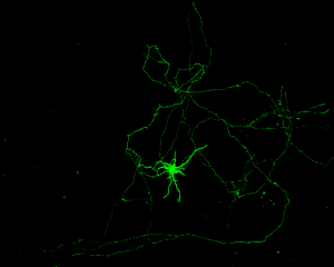
 and MAP2 (green). The neurosphere is constantly refining its network throughout the differentiation process just like J.A.R.V.I.S. (Just A Rather Very Intelligent System).")

 cells on a substrate coated with collagen nanofibers. The collagen nanofibers have been produced with a template-assisted extrusion process under physiological conditions.")

 colony
undergoing neural induction to form neural progenitor cells (NPCs).
Immunofluorescence staining was done at day 7 of neural induction using a 2D differentiation protocol. Here, standard NPC markers nestin (green) and Sox1 (red) along with DAPI (blue) are visualized to highlight that a neural identity is present after 7 days of neural differentiation from a pure iPSC reprogrammed initially from renal epithelial cells found in urine.")

 and in the background the trabeculae lining the left ventricle.")
 closely associated with perfusable vessels (green) when transplanted underneath the skin. Engineered pseudo-islets are being explored as an approach to treat Type 1 diabetes.")


, stained with DAPI (blue), Sox1 (red), and nestin (green), can differentiate into neurons that inhabit our heads and give us our intelligence. The purity of NPC population in a given culture is the key to successful investigation of neurological
disorders.")

work\" by Krystal Jacques-Smith. A clonal pancreatic sphere isolated from EII PDX1-Cre xRosa YFP mouse. The sphere contains hundreds to thousands of pancreatic stem cell-derived progenitors that have differentiated on Matrigel into insulin positive cells marked as red vesicles surrounding the DAPI labelled nucleus of some cells. Ubiquitous yellow in the sphere indicate that its progeny come from the PDX1 pancreatic lineage.")
 stained for nestin (blue), vimetin (magenta) and DAPI (blue). This work reflects the hetergenous and dynamic phenotypes displayed by MSC populations during culture. Culture-dependent establishment of MSC populations is paradoxical and routinely require in-depth phenotypic and functional characterizations.")

. Cells are stained with MAPs (red), Sox2 (green) and Hoechst (blue).")
, they wanted to show that reciprocated love.")

 and nuclear architecture (magenta).")
 and PDGFRa Cre tdTomato (red).")
-expressing pancreatic beta-like cells, derived from stem cells. These “spheroids” of beta-like cells are being explored as a possible avenue of therapeutic treatment for type 1 diabetic patients, who have lost their pancreatic beta cells due to autoimmune attack.")


, derived from H9-hESC, were stained with NSC marker nestin, which is green in colour, and nucleus stained with DAPI,
red in colour.")
.")